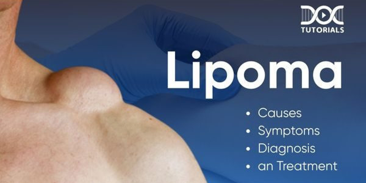

Diagnostic Technologies for Lipoma Assessment in Riyadh
Lipoma Treatment in Riyadh has reached a high level of clinical sophistication, largely due to the integration of advanced diagnostic technologies that allow for precise identification and treatment planning. Before any surgical or non-surgical intervention is considered, the diagnostic phase is crucial to differentiate a common lipoma from other subcutaneous lesions. By utilizing state-of-the-art imaging and diagnostic tools, medical professionals in the city can confidently assess the nature, depth, and anatomical relationship of these fatty growths, ensuring that the chosen treatment path is the most safe and effective for the individual patient.
Clinical Evaluation: The First Step
The diagnostic process begins with a detailed physical examination. A skilled clinician will palpate the lesion to assess its consistency, mobility, and boundaries. Lipomas typically feel soft, rubbery, and mobile, moving slightly under the skin when pressure is applied. During this stage, the physician will also gather a medical history, looking for signs such as rapid growth, associated pain, or a history of similar lumps, which helps in ruling out less common conditions like liposarcoma or other soft-tissue tumors.
The Role of Ultrasound Imaging
Ultrasound remains the frontline diagnostic tool for Lipoma Treatment in Riyadh due to its safety, availability, and high efficacy.
High-Resolution Imaging: Modern ultrasound machines provide clear, real-time images that allow the practitioner to differentiate between a simple lipoma, a fluid-filled cyst, or a more complex vascular mass.
Defining Borders: Ultrasound helps determine the boundaries of the lipoma, which is vital for surgical planning. It can reveal if the mass is superficial or if it extends into deeper muscle layers.
Lack of Radiation: As an imaging technique that utilizes sound waves rather than ionizing radiation, ultrasound is an ideal, non-invasive method for monitoring lumps over time, especially in patients who prefer a "watch and wait" strategy before deciding on removal.
Advanced Imaging: MRI and CT Scans
While ultrasound is excellent for superficial lumps, larger or deeper lipomas may require more comprehensive imaging to ensure surgical safety.
Magnetic Resonance Imaging (MRI): MRI is considered the "gold standard" for complex cases. It provides superior soft-tissue contrast, allowing the surgeon to see exactly how the lipoma relates to nearby nerves, major blood vessels, and fascia. This level of detail is particularly important for lipomas located in delicate areas such as the neck, axilla, or limbs where critical anatomical structures are present.
Computed Tomography (CT): Although less common for routine lipoma assessment, a CT scan may be used if the healthcare team needs to assess the density of the mass more accurately or if an MRI is contraindicated for the patient.
Biopsy and Histopathological Examination
If a clinical examination or imaging suggests that a lump has "atypical" features—such as hardness, fixity to surrounding tissue, or an unusually rapid growth rate—a biopsy may be performed.
Fine Needle Aspiration (FNA): A thin needle is used to extract a small amount of tissue for analysis.
Core Needle Biopsy: In some cases, a larger sample is needed to provide a definitive diagnosis.
Histopathology: Once the tissue is extracted, it is examined by a pathologist to confirm that the cells are benign fat cells. This analysis is the final word in confirming a diagnosis and ensures that any treatment plan is based on an accurate understanding of the tissue’s nature.
The Impact of AI-Driven Diagnostics
Looking toward the future of care in 2026, many diagnostic centers in Riyadh are beginning to integrate artificial intelligence into their workflow. AI algorithms are now being utilized to analyze ultrasound and MRI imagery with high precision. By comparing patient data against massive databases of known soft-tissue tumors, these systems can assist clinicians in identifying potential risks or characteristics that might have been overlooked by the human eye alone. This technological support enhances the safety of the diagnostic process and leads to more personalized, data-backed treatment recommendations.
Informed Decision Making
The goal of this comprehensive diagnostic approach is to empower the patient with clear information. By clearly understanding what the lump is, where it sits, and how it interacts with the surrounding anatomy, patients in Riyadh can make informed choices about their treatment. Whether the result is a recommendation for simple observation or a plan for surgical excision, the use of these advanced diagnostic tools ensures that every patient receives a high standard of care tailored to their specific medical needs.