
Cryotherapy: A Fast and Effective Method for Skin Lesion Removal in Riyadh
Opting for advanced clinical cold therapy is a highly popular pathway for individuals seeking swift Skin Lesion Removal in Riyadh without the downtime or complexity of surgical incisions. Riyadh's leading dermatological centers widely utilize cryotherapy as a foundational non-surgical modality to instantly eliminate a diverse range of superficial skin irregularities. Known colloquially as "lesion freezing," this medical technique leverages the extreme temperatures of pressurized liquid nitrogen to target and safely destroy compromised surface cells. Because the method requires no incisions, no surgical stitching, and often zero local injected anesthesia, it has established itself as an incredibly safe, efficient, and cost-effective outpatient solution for achieving clear skin.
The Scientific Principles Behind Cryo-Necrosis
Cryotherapy functions by inducing targeted, controlled cryo-necrosis (cellular destruction via extreme cold). The specialized medical delivery systems utilized in modern clinics house liquid nitrogen, which is maintained at a blistering temperature of -196 degrees Celsius.
When this ultra-cold element is administered to an unwanted skin growth, it initiates an immediate multi-phase biological reaction. First, the water content both inside and directly surrounding the abnormal skin cells instantly solidifies into sharp micro-ice crystals. As these ice crystals expand, they physically puncture and tear the cellular membranes of the lesion. Simultaneously, the extreme cold causes localized micro-blood vessels supplying the growth to constrict and shut down, cutting off the oxygen supply to the targeted tissue. Once the freezing cycle concludes and the area begins to thaw, the disrupted cells collapse, die, and are naturally rejected by the body’s immune system, clearing the path for fresh epidermal tissue to regenerate.
What Conditions Can Be Safely Treated?
While cryotherapy is an exceptionally versatile tool, it is specifically engineered to treat superficial lesions that reside primarily within the outermost layers of the skin. A specialist will thoroughly examine your skin structure to determine if your specific growth is an ideal candidate.
Viral Warts and Verrucas: Cryotherapy boasts incredibly high success rates (often between 75% and 98%) in clearing stubborn hand, finger, and foot warts by freezing out the virally infected tissue layers.
Acrochordons (Skin Tags): Small, hanging stalks of skin that form in high-friction areas like the neckline or armpits can be frozen at their base, causing them to drop off effortlessly within a few days.
Seborrheic Keratoses: These benign, waxy, or crusty brown growths that frequently develop as a result of natural skin maturation respond beautifully to localized cold therapy.
Actinic Keratoses: These rough, sandpapery, scaly patches are caused by chronic ultraviolet exposure from the sun. Because they are classified as precancerous, removing them via cryotherapy is a vital preventative healthcare measure to stop them from mutating into skin cancer.
The Swift In-Clinic Outpatient Experience
One of the greatest practical advantages of undergoing clinical cryotherapy is the sheer speed and simplicity of the physical appointment. The entire removal process is treated as a brief, walked-in outpatient service that rarely takes more than 10 to 15 minutes to complete.
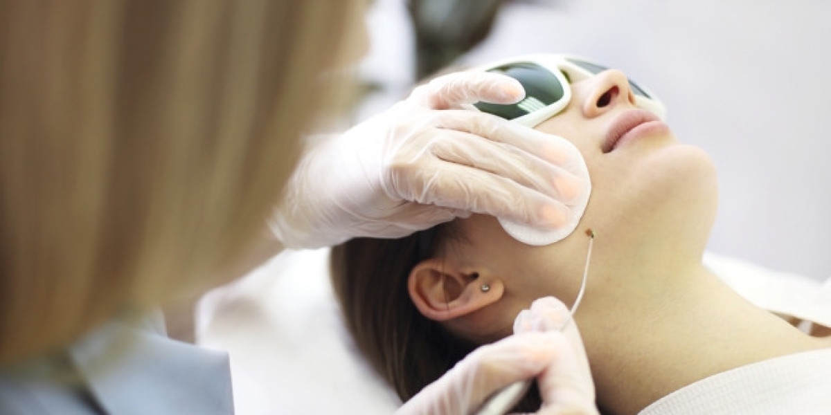
The treatment area is first cleansed thoroughly with a specialized medical antiseptic. Because the procedure is completely non-invasive and does not involve cutting the skin, local numbing injections are typically unnecessary; the extreme cold itself acts as a natural local anesthetic. The clinician uses a specialized, hand-held spray canister equipped with a micro-diameter nozzle to deliver a highly focused blast of liquid nitrogen directly onto the lesion for a controlled window usually lasting between 5 to 20 seconds. During the application, you will feel a sharp, intensely cold biting or stinging sensation, followed by a localized throbbing feeling as the tissue begins to thaw. Once the brief application is finished, a soothing protective ointment is applied, and you can comfortably drive yourself home and resume your entire daily schedule immediately.
Understanding the Step-by-Step Healing Timeline
Because cryotherapy deliberately alters the target tissue to stimulate cellular shedding, the treated site undergoes a distinct, predictable transformation over the course of a few weeks. Knowing what to expect stage-by-stage ensures a stress-free recovery.
[Day 1-2: Redness & Swelling] -> [Day 2-5: Mild Blister Forms] -> [Day 5-10: Crusting/Scabbing] -> [Day 10-21: Natural Shedding]Within the first 24 to 48 hours following your session, the treated area will develop minor redness and localized swelling, mimicking a small burn. Between days two and five, a small, fluid-filled blister will naturally develop directly beneath or within the frozen lesion. This blister is a healthy sign that the dead cells are separating cleanly from the underlying dermis.
By the end of the first week, the blister will naturally dry out, transitioning into a flat, dark crust or scab. Over the subsequent ten days, this protective crust will gradually flake away entirely on its own, revealing smooth, fresh, slightly pink skin beneath. The entire structural cycle from initial freezing to final shedding is typically completed within two to three weeks.
Rules for Safe Aftercare and Optimal Aesthetics
Achieving a clean cosmetic outcome with zero residual marking relies heavily on protecting the delicate healing site at home. By adhering to a few basic clinical skin rules, you can ensure your skin blends back together flawlessly:
Never Force the Scab: It is absolutely imperative that you never scratch, pick, or prematurely peel away the drying scab or pop the fluid blister. Forcing the crust off exposes raw, unhealed tissue to external bacteria and dramatically increases your risk of developing a permanently pitted or darkened scar.
Practice Moist Healing: Keep the site hydrated by applying a thin dab of pure petroleum jelly or an approved recovery ointment daily. Hydrated wounds allow new skin cells to travel across the surface significantly faster, preventing thick, jagged textures from forming.
Defend Against the Sun: Freshly uncovered pink skin cells are highly susceptible to UV damage. Apply a broad-spectrum sunscreen with an SPF of 50 or higher to the site daily to prevent post-inflammatory hyperpigmentation, allowing the temporary pink mark to fade smoothly into your natural skin tone.